Severe and Extensive Bite Wound Treatment
PROBLEM LIST/ DIFFERENTIAL DIAGNOSIS
Traumatic wound with large deficit Cardiovascular compromise. Risk of sepsis without prompt treatment.
PRE-OPERATIVE MANAGEMENT
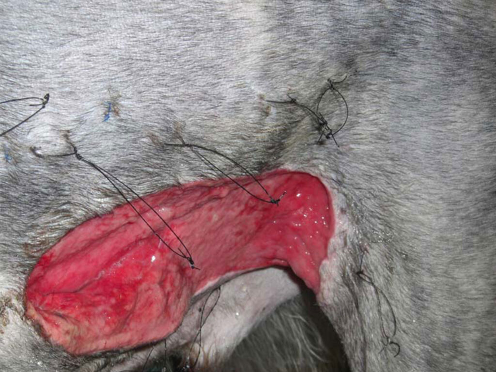
Fluid therapy was initiated using ringer’s lactate solution at 90ml/kg/hr for the first 15 min then 10ml/kg/hr. Clavulanic/Amoxicillin antibiotics were given 20mg/kg IV and the wound was protected temporarily with a dressing. After an hour the dog was sedated for a close inspection, cleaning debridement, and irrigation of the wound. The large skin flap was sutured in place trying to preserve as much skin as possible. After several days the true extent of the injury was evident and the wound was managed as an open wound with initial surgical debridement and daily irrigation. Enterococcus caecofaecalis was cultured from this wound. The wound was managed until negative culture and no necrosis was seen. The antibiotic was replaced with Enrofloxacin and Metronidazole based on culture and sensitivity. The wound size was reduced substantially and a healthy granulation bed was evident.

PRESENTATION & HISTORY
A three-year-old male neutered Irish Wolf Hound weighing 64kg presented with a severe and large bite wound on the left flank and left abdominal area. The wound measured 40×15 cm across the left abdomen including some of the inguinal area and the craniomedial thigh. The dog was bitten by a
Grey hound while in the park and was presented to our veterinary practice an hour later.
POST-OPERATIVE OUTCOME/COMPLICATIONS
When the wound was examined early the following week it was evident that most of it healed in an
unusual rate and that the pre planned skin flap would not be needed any
more and that this wound would continue to heal as an open wound.
CLINICAL EXAMINATION & INVESTIGATION
The dog was distressed, panting, and restless. He had slightly pale mm and tachycardia. The wound appeared fresh and very traumatic with a large tissue deficit. A large degloving injury emerged and it was unclear as to the future of this large flap of skin. No other pathologies were noted apart of the large wound.
PROBLEM LIST/ DIFFERENTIAL DIAGNOSIS
Traumatic wound with large deficit Cardiovascular compromise
Risk of sepsis without prompt treatment.
PRE-OPERATIVE MANAGEMENT
Fluid therapy was initiated using ringer’s lactate solution at 90ml/kg/hr for the first 15 min then 10ml/kg/hr. Clavulanic/Amoxicillin antibiotics were given 20mg/kg IV and the wound was protected temporarily with a dressing. After an hour the dog was sedated for a close inspection, cleaning debridement, and irrigation of the wound. The large skin flap was suture in place trying to preserve as much skin as possible.
After several days the true extent of the injury was evident and the wound was managed as open wound with initial surgical debridement and daily irrigation. Enterococus caecofaecalis was cultured from this wound.
The wound was managed until negative culture and no necrosis was seen. The antibiotic was replaced with Enrofloxacin and Metronidazole based on culture and sensitivity. The wound size was reduced substantially and healthy granulation bed was evident.
PRE-OPERATIVE MANAGEMENT
Fluid therapy was initiated using ringer’s lactate solution at 90ml/kg/hr for the first 15
min then 10ml/kg/hr. Clavulanic/Amoxicilin anibiotic was given 20mg/kg IV and the wound was protected temporarily with a dressing. After an hour the dog was sedated for a close inspection, cleaning debridement and irrigation of the wound. The large skin flap was suture in place trying to preserve as much skin as possible. After several
days the true extent of the injury was evident and the wound was managed as open wound with initial surgical debridement and daily irrigation. Enterococcus caec of aecalis was cultured from this wound. The wound was managed until negative culture and no necrosis was seen. The antibiotic was replaced with Enrofloxacin and Metronidazole based on culture and sensitivity. The wound size was reduced substantially and a healthy granulation bed was evident.


SURGICAL PROCEDURE
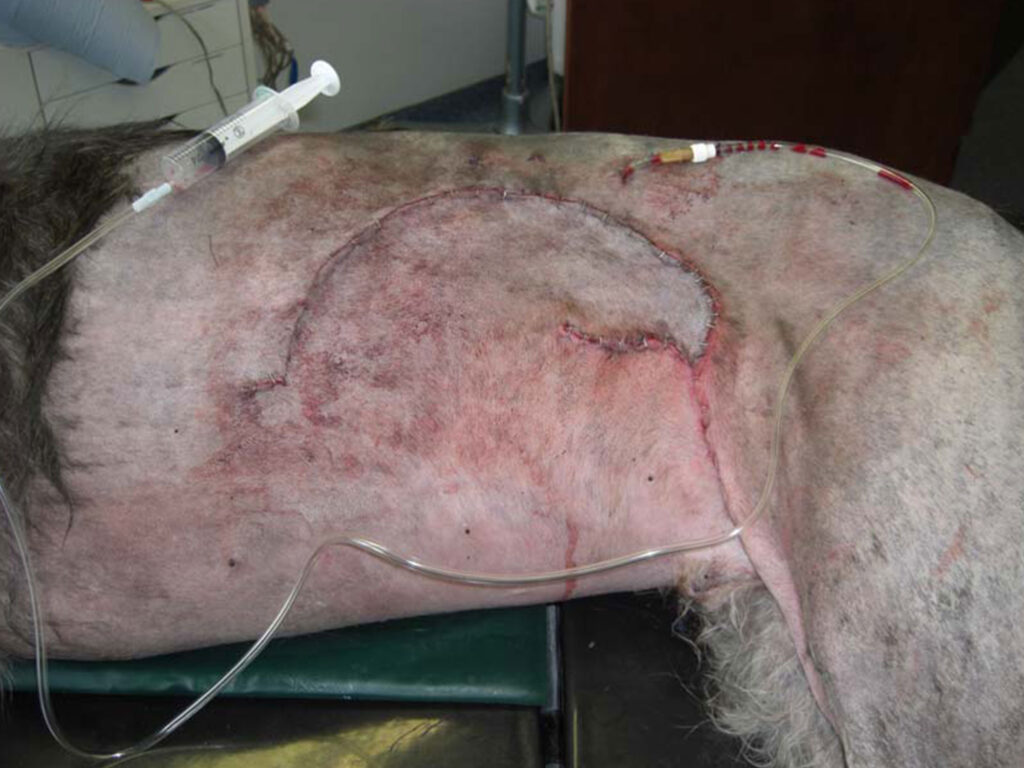
Ten days post initial presentation the wound bed allowed closure. The vast skin deficit
was closed using a sub-dermal plexus rotational skin flap.
POST-OPERATIVE CARE
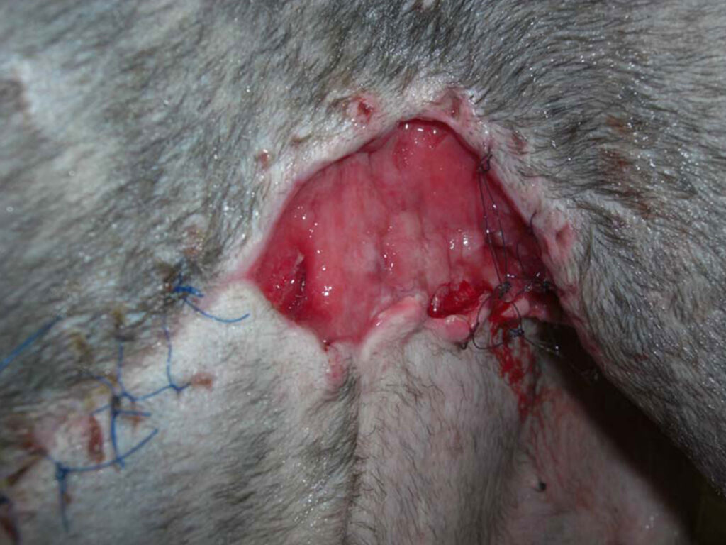
The distal aspect of the flap dehisced three days post-surgery, and the flank area became open, due to excessive motion and self-mutilation. This was managed as a
pen wound with regular irrigations and VetGold for a few days. A second delayed closure was planned for the following week, utilizing an axial pattern flap from the caudal superficial epigastric artery. VetGold was continued TID over the weekend and the wound was protected from self-trauma.
POST-OPERATIVE OUTCOME/COMPLICATIONS
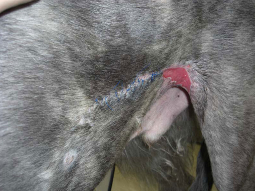
When the wound was examined early the following week it was evident that most of it
healed at an unusual rate and that the pre-planned skin flap would not be needed any
more and that this wound would continue to heal as an open wound.

FOLLOW-UP
The dog was discharged with VetGold and applied BID. The wound healed completely after two weeks.
DISCUSSION
Bite wounds are often misleading when presented shortly following the incident. The nature of this injury is very traumatic than hiding vast tissue compromise and infection. It is well established that these wounds should be dealt with open until the true extent of the lesion is determined.
In most cases of these large wounds, infection would be present and need to be dealt with before final closure. In this case, the large skin flap did not survive despite its initial healthy appearance. Enterococcus infection was present most likely harbored in the attacking dog’s teeth.
The last 10-15% of the skin flap used to close the ready wound, was dehisced mostly due to movement at the flank area and self-mutilation as the dog managed to reach the wound despite its protection. The distal part of a sub-dermal plexus flap is the most vulnerable and proved to be unreliable in this case.
The edge of the flap became necrotic and was cleaned. VetGold was applied for about a week until the wound was inspected prior to a second skin flap for closure. Due to the rapid wound contraction and healthy granulation bed, it was decided to allow the wound to heal as an open wound. This would reduce costs substantially, eliminate the necessity of anesthesia, and reduce total hospitalization time.